







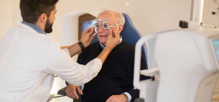
Maintaining quality of life, personal dignity, and day-to-day independence during the senior years depends heavily on preserving healthy visual function. Simple daily activities—such as reading prescription medication labels, preparing nutritious meals, navigating staircases, managing household finances, and driving safely—rely on sharp, clear eyesight. However, as human anatomy matures, the delicate structures of the eye become increasingly vulnerable to progressive, sight-threatening conditions. Navigating senior vision care requires a proactive approach that combines routine clinical screenings, early medical intervention, and a clear understanding of provincial health coverage frameworks.
Scheduling an annual, comprehensive eye exam provides the absolute first line of defence against progressive, irreversible vision loss. Across Canadian provinces, senior healthcare frameworks generally fund or heavily subsidize annual ocular assessments for older adults, ensuring that preventive care remains accessible to aging populations. When early signs of ocular disease emerge, your eye doctor collaborates directly with your family doctor to manage underlying systemic factors like diabetes, hypertension, and systemic inflammation, keeping your vision clear and supporting your overall health throughout your golden years.
Major Age-Related Ocular Conditions: Early Signs and Pathology
Age-related eye diseases often develop silently over several years. They cause permanent damage to delicate neural tissue long before noticeable visual symptoms appear. Understanding the underlying pathology and primary warning signs helps seniors, family members, and caregivers seek timely clinical care before functional sight is lost.
Age-Related Macular Degeneration (AMD) stands as a leading cause of severe visual impairment among adults aged sixty and older. This condition impacts the macula, which is the specialized central portion of the retina. The macula provides the sharp, straight-ahead vision required for reading and recognizing faces. AMD occurs in two distinct clinical forms: dry AMD and wet AMD.
Dry AMD accounts for the vast majority of cases. It progresses gradually as light-sensitive cells in the macula break down over time. Small, yellow protein deposits called drusen build up beneath the retinal pigment epithelium. Over time, geographical atrophy develops, causing central blurry spots and distorted vision.
Conversely, wet AMD is less common but significantly more aggressive. It occurs when abnormal, fragile blood vessels grow beneath the retina in a process known as choroidal neovascularization. These delicate vessels frequently leak blood, lipids, and fluid into macular layers, causing sudden swelling and rapid central vision loss. Early detection through advanced retinal imaging is essential. Modern anti-VEGF injections can stop vessel growth and preserve functional sight if administered promptly.
- Cataracts: A natural clouding of the eye’s crystalline lens caused by oxidative protein clumping, leading to faded colours, poor night vision, halos around streetlights, and persistent glare.
- Glaucoma: Often called the “silent thief of sight,” this group of eye conditions damages the optic nerve, typically due to elevated intraocular pressure caused by poor fluid drainage inside the anterior chamber.
- Diabetic Retinopathy: Microvascular retinal damage caused by chronically elevated blood sugar levels, which weakens capillary walls, causes fluid leakage, and sparks abnormal vessel growth.
Silent Threats: Glaucoma and Diabetic Eye Disease
Glaucoma poses a unique diagnostic challenge. Open-angle glaucoma, the most common form, causes zero physical pain and no early visual symptoms. The condition develops when the eye’s natural drainage pathway, the trabecular meshwork, becomes clogged or inefficient over time. Fluid then builds up inside the front of the eye, raising intraocular pressure. This pressure places harmful mechanical stress on the delicate nerve fibres forming the optic nerve.
Because glaucoma destroys peripheral vision first, patients rarely notice vision loss early on. Significant, permanent damage often spreads toward the center of the visual field before symptoms appear. By the time a senior notices missing patches in their side vision, thousands of optic nerve fibres are permanently lost. Regular pressure checks and nerve head scans during annual checkups remain the only reliable way to catch glaucoma early and start pressure-lowering drop therapies.
Diabetic retinopathy similarly threatens senior vision through silent microvascular destruction. Chronically high blood sugar levels damage the tiny capillaries that nourish the retina. These damaged vessels leak fluid, protein, and blood directly into retinal tissue. In advanced stages, known as proliferative diabetic retinopathy, the retina suffers from severe oxygen deprivation. It releases chemical signals that trigger the growth of fragile new blood vessels. These delicate vessels bleed easily into the vitreous humour and create scar tissue that can pull the retina away from the back of the eye. This risk makes strict blood sugar control and annual dilated eye exams essential for all diabetic seniors.
Navigating Canada’s Senior Healthcare Framework and Diagnostic Tools
Recognizing the vital role that clear sight plays in senior health and falls prevention, Canadian provincial healthcare plans prioritize routine vision care for older demographics. Provincial coverage across most regions funds routine annual eye assessments for adults aged sixty-five and older. These covered visits remove financial barriers, allowing seniors to undergo comprehensive evaluations without out-of-pocket stress.
Modern optometric clinics utilize sophisticated, non-invasive diagnostic technologies to monitor ocular health with microscopic precision over time. Optical Coherence Tomography (OCT) uses light waves to capture cross-sectional 3D images of the retina and optic nerve head, allowing practitioners to map retinal layers down to the micrometre and spot cellular swelling long before it shows up on standard visual tests.
Digital fundus photography takes high-resolution images of the back of the eye, creating a permanent visual record that clinicians compare year after year to track subtle vessel changes, drusen growth, or optic disc cupping. Additionally, computerized visual field testing maps peripheral sensitivity, ensuring that subtle blind spots caused by glaucoma or neurological events like strokes are identified and treated early.
Collaborative Medicine: Connecting Optometry and Primary Care
Managing senior health effectively requires a coordinated, multidisciplinary approach where primary care physicians and ocular specialists work in close partnership. The eye does not exist in isolation, and structural changes observed within the retina frequently reflect broader systemic health challenges.
For instance, when an eye specialist detects microaneurysms, cotton wool spots, or retinal hemorrhages during a routine checkup, they immediately communicate these findings to the patient’s primary care provider. This feedback allows the primary care physician to adjust diabetes medications, reassess blood pressure management, or order comprehensive cardiovascular lab work.
Likewise, when primary care doctors manage patients with autoimmune diseases like rheumatoid arthritis, thyroid conditions, or vascular disease, they coordinate regular eye monitoring to catch secondary complications early. This collaborative healthcare loop ensures that seniors receive holistic care that protects both their vision and their overall physical vitality.
Proactive Strategies for Long-Term Visual Independence
Clinical care provides the baseline for managing age-related eye conditions, but daily lifestyle habits play a major supporting role. Wearing broad-spectrum UV-blocking sunglasses protects your eyes from ultraviolet rays. This simple step helps delay cataract formation and reduces oxidative stress on your retina.
A balanced diet rich in leafy greens, colourful vegetables, omega-3 fatty acids, and key antioxidants nourishes delicate eye tissues and supports macular health. Dietary discipline, regular exercise, and prescribed medications help manage chronic conditions like high blood pressure, high cholesterol, and diabetes. Keeping these health factors under control ensures your retinal blood vessels stay healthy and resilient.
Taking advantage of provincial senior care coverage, scheduling annual checkups, and keeping open lines of communication with your care team ensures eye diseases are caught early. Addressing changes at the earliest stage makes managing them far more effective. Prioritizing your eye health preserves your independence, protects your quality of life, and keeps you enjoying your favourite activities throughout your senior years.