The modern game of golf has expanded beyond traditional outdoor courses into highly immersive digital environments. The indoor golf simulator has become a central training and recreation tool for players seeking year-round consistency and data-driven improvement. These systems rely on fast-processing sensors, high-resolution visuals, and real-time feedback that translate physical movement into detailed performance metrics. Because so much of this experience depends on visual interpretation, clarity of sight becomes an important factor in how effectively a player engages with the simulation.

Visual Demands of the Indoor Golf Simulator

A modern indoor golf simulator is designed to replicate real-world golfing conditions through a combination of projection systems, tracking cameras, and analytical software. It renders ball flight, spin, launch angle, and course layout with a level of detail that requires rapid visual processing. Unlike outdoor play, where natural depth cues and environmental context assist perception, simulator environments compress all information into a controlled visual interface.

Players must interpret layered data such as trajectory arcs, landing predictions, and terrain gradients, often presented simultaneously on screen. Even small delays in visual interpretation can affect how a player understands shot quality or makes adjustments between swings. As a result, visual clarity becomes not just helpful but essential for extracting meaningful insight from each session.

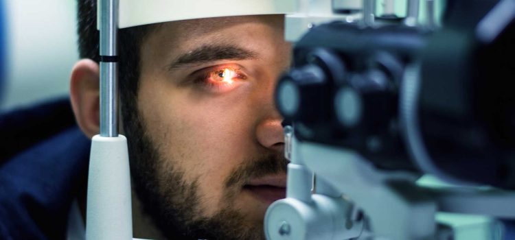
The Role of the Optometrist in Visual Interpretation

Within performance-based simulation environments, the optometrist is often associated with maintaining the clarity and precision of visual input. While the focus is not on treatment or correction in this context, structured visual evaluation helps ensure that players can engage fully with detailed digital feedback systems.

The indoor golf simulator environment places unique demands on visual coordination. Users must shift attention between near-field data displays and the simulated perception of distance and depth on screen. This requires the visual system to manage contrast sensitivity, motion tracking, and focus transitions at a rapid pace.

When these visual elements are not well aligned with the demands of the simulator interface, interpretation can become less efficient.

Translating Visual Clarity into Real-Time Performance Data

One of the defining strengths of the indoor golf simulator is its ability to convert physical motion into immediate, highly detailed feedback. Each swing generates a stream of data points, including club speed, face angle, launch direction, and carry distance. These metrics are often displayed alongside a visual representation of the shot, allowing players to compare intent with outcome.

For this feedback loop to be effective, the user must be able to quickly and accurately connect visual cues with numerical results. Sharp visual interpretation helps reduce the cognitive delay between seeing a result and understanding its meaning. This is especially important when making incremental adjustments between swings.

In this environment, clarity of vision supports the overall learning process. The simulator provides the data, but the player must visually integrate that data into actionable understanding.

Maintaining Consistency Across Digital Practice Sessions

Consistency is a key objective in both traditional golf and simulator-based training. The indoor golf simulator offers controlled conditions that eliminate external variables such as wind, uneven terrain, or weather changes. However, the visual environment itself can still vary depending on screen brightness, projection quality, and session duration.

Over time, extended exposure to detailed visual interfaces can place continuous demands on focus and attention. Maintaining stable visual interpretation helps ensure that performance trends reflect true skill development rather than fluctuations in perception.

The optometrist plays a key role in supporting visual accuracy. While often associated with routine eye examinations, their relevance extends into any activity that depends on fast, precise visual interpretation. The goal is to help ensure that the visual information being presented by the simulator is perceived as clearly and consistently as possible.

As golf continues to evolve through technology, the indoor golf simulator has become a powerful tool for analysis, repetition, and skill refinement. Its effectiveness depends heavily on the player’s ability to interpret dense visual data and respond in real time. Within this framework, the role of the optometrist is connected to the broader importance of visual clarity in high-performance environments.

Ultimately, success in simulator-based training is not only about swing mechanics or equipment familiarity, but also about how clearly and efficiently the visual system processes information.

When most people think about scheduling a vision checkup, they view the appointment through a very narrow lens. They assume that the primary goal of the visit is simply to determine whether their vision has changed or if they need a new prescription for glasses or contact lenses. If they can read street signs clearly and do not experience frequent blurry vision while working on their computers, they often skip these visits entirely for several years. This common oversight stems from a basic misunderstanding of what a comprehensive vision evaluation actually checks.

The human eye is one of the most complex, interconnected organ systems in the entire body. It is the only place on the human anatomy where a medical professional can view living blood vessels, active nerve tissues, and microscopic connective fibres directly, without making a single surgical incision. Because of this structural access, a comprehensive vision checkup serves as a powerful diagnostic tool for your entire physical health. Booking a routine eye exam allows an optometrist to spot the very first warning signs of serious, systemic medical conditions long before any physical symptoms show up in your daily life. Sharing these ocular findings with your family doctor allows your primary care team to step in early, completely changing how you manage long-term chronic illnesses.

The Vascular Connection: Spotting High Blood Pressure and Diabetes

The delicate blood vessels running across the back of your retina are highly sensitive to changes in your circulatory system. When an individual develops chronic high blood pressure, the immense force of the blood pumping through the body damages these tiny arteries first. During a dilated eye examination, an optometrist can see these vessels narrowing, twisting, or even leaking small amounts of blood into the surrounding retinal tissue. This condition, known as hypertensive retinopathy, often occurs long before a patient ever experiences a headache or registers a high reading on a standard blood pressure cuff.

Similarly, the eyes are one of the primary target areas for tracking the progression of diabetes. High blood sugar levels actively weaken the structural walls of the microscopic blood vessels in the retina. This creates a critical retinal vascular diagnostic pathway where elevated systemic pressure or blood sugar damages fragile vessels first. These visible retinal leaks and arterial narrowings are then caught during a routine dilated eye screening, allowing the optometric report to be shared with primary care. This critical data allows for fast medical intervention, helping ensure early disease management and long-term vision preservation. Because these early vascular changes occur on a microscopic scale at the back of the eye, they do not cause any immediate pain or vision loss. Catching these early leaks allows your primary physician to alter your diet or update your medication quickly, protecting both your vision and your cardiovascular health from irreversible damage.

Detecting High Cholesterol and Neurological Risks Early

Beyond tracking blood pressure and blood sugar, a professional eye evaluation reveals hidden indicators of dangerous cholesterol and neurological conditions. When low-density cholesterol accumulates in the bloodstream, fatty deposits settle inside the eye’s structures, showing up as arcus senilis—a distinct blue or white ring around the outer edge of the cornea. This condition provides a clear marker for elevated cholesterol, while deeper within the eye, the retina can display a Hollenhorst plaque, a bright yellow cholesterol crystal indicating urgent carotid artery blockage risks. Additionally, the optic nerve disk can develop papilledema, where distinct structural swelling serves as an immediate warning sign of dangerous intracranial pressure.

Because the optic nerve provides a direct physical link between the eye and the central nervous system, it functions as an invaluable tool for tracking brain health. The optic nerve head sits at the back of the eye, acting as a direct data cable that sends visual signals straight to the brain cavity. If a patient develops an underlying condition that increases pressure inside the skull—such as a brain tumor, aneurysm, or severe inflammation—that pressure travels down the nerve path and forces the disk to swell. An eye doctor can spot this swelling instantly during a routine exam, allowing them to refer the patient for emergency neurological imaging immediately and stopping life-threatening issues before they cause catastrophic damage.

A Balanced Routine Checklist for Total Physical Wellness

Protecting your total physical health requires consistent attention and regular preventative checkups. Treating your vision care as a major element of your primary healthcare planning ensures that your medical team catches hidden problems early. Use this simple wellness checklist to coordinate your annual appointments and keep your entire body functioning safely:

  • Schedule Your Regular Physical: Visit your primary care physician annually for standard blood work, health history updates, and milestone physical checkups.
  • Book Your Yearly Vision Evaluation: Ensure every member of your family receives a comprehensive eye test every twelve to twenty-four months, regardless of whether they wear glasses.
  • Request a Dilated Retinal Scan: Ask your eye care provider to perform a dilated exam or take high-resolution digital retinal photos to view the deep blood vessels clearly.
  • Share Medical Updates Across Clinics: Request that your optometrist send an official copy of your vision report straight to your primary care clinic for your permanent files.
  • Monitor Changes in Daily Vision: Alert your doctor immediately if you experience sudden flashes of light, a wave of new floaters, or frequent unexplained headaches after working.

Protecting Your Health Through Integrated Medicine

True healthcare success relies on taking a comprehensive, big-picture approach to your body’s internal systems. Your vision health is deeply connected to your cardiovascular, metabolic, and neurological well-being. Treating your annual eye examination as a vital diagnostic tool allows you to build a powerful defence system that catches dangerous chronic illnesses at the very beginning.

Using specialized local medical clinics and maintaining regular communication with your primary physician gives you the tools needed to make smart, confident choices for your family. Sharing diagnostic reports between your optometric and primary care teams creates a seamless network of protection that eliminates hidden health risks. By prioritizing your routine eye examinations today, you protect your precious sight, secure early intervention for your internal health, and enjoy total peace of mind for the years ahead.